Draw an Ecg Waveform Representing One Heartbeat
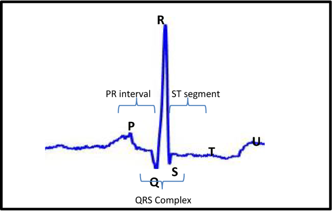
This post will walk you through the different parts of an EKG wave and how it applies to one cycle of the conduction system of the heart. A U wave is sometimes present.

17 4b Electrocardiogram And Correlation Of Ecg Waves With Systole Medicine Libretexts
THE GRAPHIC RECORDING OF THE ELECTRICAL CHANGES DEPOLARIZATION FOLLOWED BY REPOLARIZATION OCCURING DURING THE CARDIAC CYCLE.
. Our solutions are written by Chegg experts so you can be assured of the highest quality. It consists of a collection of waves which represents the ventricular depolarisation. Depolarization followed by repolarization occuring during the cardiac cycle.
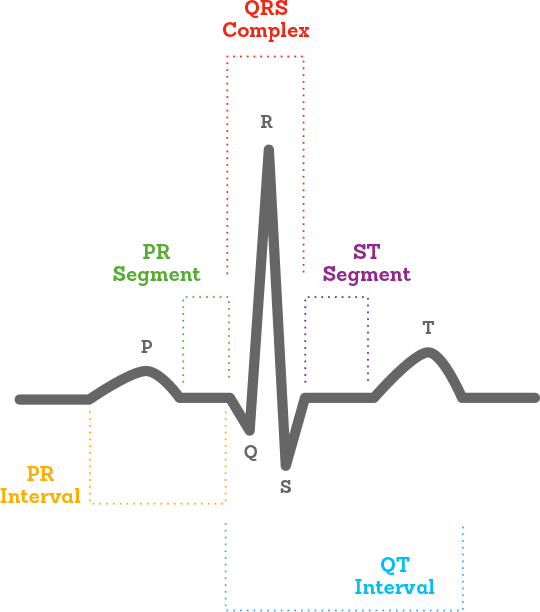
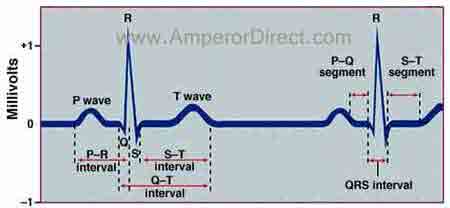
The PR interval the PR segment the ST segment and the QT interval. It leads to ventricular depolarization. Looked at separately the waves tell us whats happening in the heart at a certain time.
012 - 020 sec. Access MasteringAP -- Standalone Access Card -- for Human Anatomy Physiology Laboratory Manuals 11th Edition Chapter E31 Problem 4E solution now. The PR interval is assessed in order to determine whether impulse conduction from the atria to the ventricles.
Draw an ECG waveform representing one heartbeat. Label the PQRS and T wavws. The hearts electrical activity is represented on the monitor or ECG tracing by three basic waveforms.
LABEL THE P QRS AND T WAVES. ECG is a test that checks how your heart is functioning by measuring the electrical activity of the heart how often the heart beats heart rate and how regularly it beats heart rhythm. The graph can show the hearts rate and rhythm.
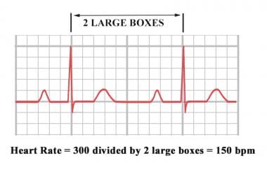
One second is marked by 5 large grid blocks thus each large block equals 02 second. THE S-T SEGMENT AND THE Q-T INTERVAL. The S-T segment.
The S-T segment and the Q-T interval. The P R and T-waves are above the base line and are known as positive waves. Graphic recording of electrical change.
Label the P QRS and T waves. Recording of electrical changes occurring during heart activity 4Draw an ECG wave form representing one heartbeat. It can present different morphologies depending on the lead read QRS complex morphology.
QT Interval QTc 040 sec Image will be uploaded soon Why is an ECG Done. Draw and label the normal EKG waveform P to U and explain each part of the wave. Depolarization of the atria the heart is in relaxation diastole contraction of the atria depolarization of the.
Label the P wave QRS complex and T wave. Between the waveforms are the following segments and intervals. Why does the heart rate increase during running.
It can also detect enlargement of the heart decreased blood flow or. As with every EZmed blog the material below will be presented simply and concisely. Together these waves represent one heartbeat.
The diagrammatic representation of a standard ECG is shown below. DRAW AN ECG WAVE FORM REPRESENTING ONE HEARTBEAT. Label the P QRS and T waves P-R interval S-T segment and Q-t interval.
Discuss how different leads represent the heart. Even though O 2 exchange is still occurring CO 2 accumulation blood pH causing cerebral vessel vasodila-tion and exciting sympathetic centers which in turn cause a reflex increase in heart rate and the respiratory drive. Analyze the ECG tracings below taking note of the heart rate rhythm and the presence and form of ECG waves.
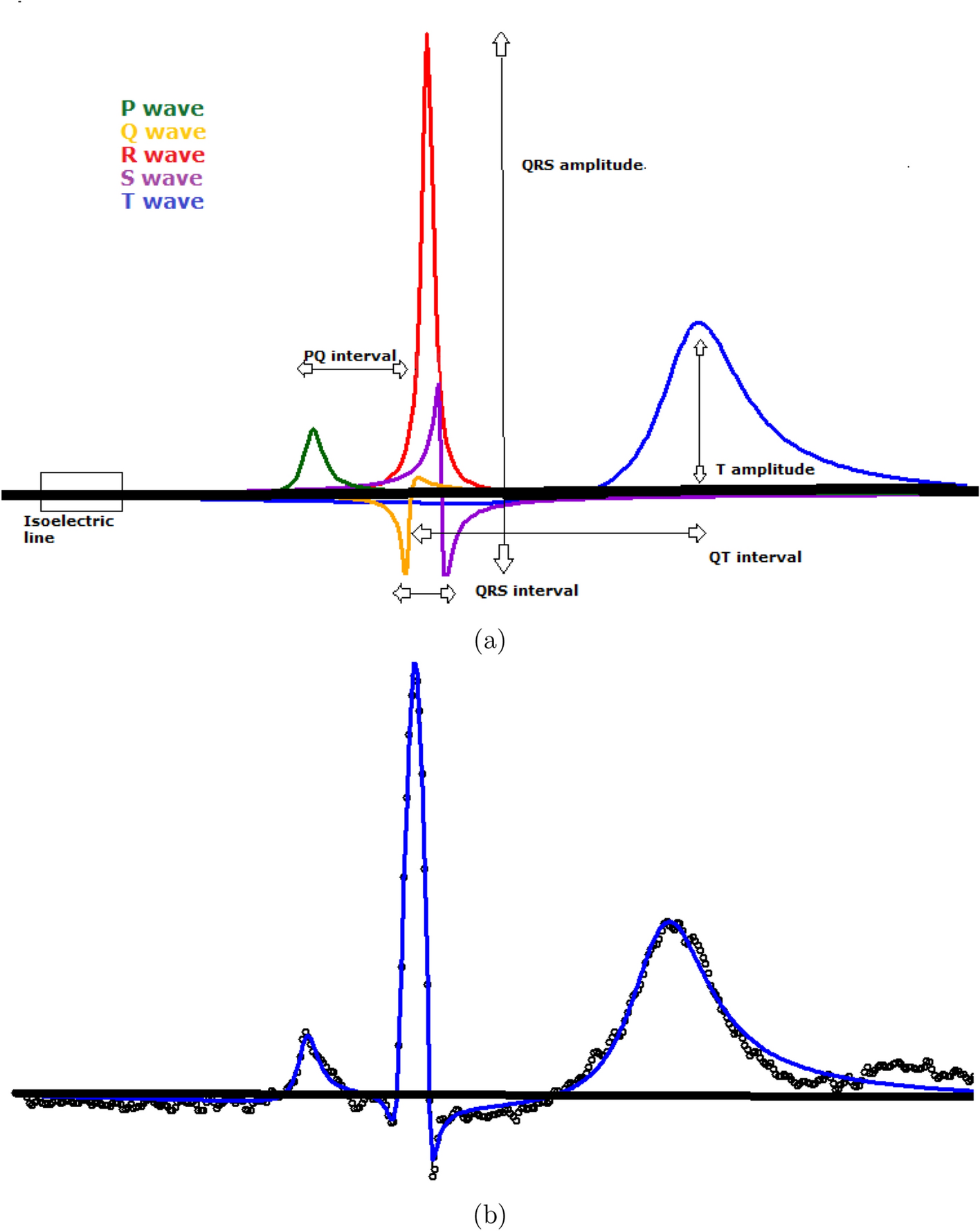
The main components of an EKG wave include the P wave PR segment QRS complex ST segment T wave and TP segment. The S-T segment and the Q-T interval. Draw an ECG wave form repping 1 heartbeat.
Feature of Normal ECG Heart beat should be in between 60-90 beats per minute bpm. The S-T segment and the O-T interval. In greater demand when our body is under strain heart has to beat faster and harder.
It represents the spread of the impulse of contraction from the AV node to the wall of the ventricle. Draw an ECG wave form representing one heartbeat. In adults ligament there now.
Outline 9 steps in interpreting the EKG. The s-t segment and the q-t interval. The S-T segment and the Q-T interval.
The P-wave PR interval and PR segment. Label the P QRS and T waves. Its duration ranges from 006 s and 010 s.
Explain placement of electrodes for 12-lead 5-lead and 3-lead EKGs. Exercise raises heart rate by acting through the sympathetic nervous system. Within the large blocks are 5 small blocks each representing 004 seconds.
The first wave is. 1 ligamentum arteriosum- called ductus arteriosus in fetal heart located between pulmonary trunk and aortic arch. Label the p qrs and t waves.
Draw an ECG wave from representing one heartbeat. C The RS wave represents the ventricular contraction of about 03 sec. Why does heart rate increase during running.
It is the first positive wave in the QRS. Draw an ECG wave form representing one heartbeat Label the P wave QRS complex and T wave. These electrodes can detect every minute form of changes happening in heart muscles and draw every depolarising pattern of heartbeat on a graph.
2 fossa ovalis - called foramen ovale in fetal heart located on right atrium wall also wall of right ventricle. The P-wave reflects atrial depolarization activation. The P wave the QRS complex and the T wave.
ECG interpretation traditionally starts with an assessment of the P-wave. Draw an ecg wave form representing one heartbeat. Electrocardiogram is a graphical representation of the cardiac cycle produced by an electrograph.
An Electrocardiogram ECG is a recording of the hearts electrical activity as a graph over a period of time as detected by electrodes attached to the outer surface of the skin and recorded by a device external to the body. 006 - 010 sec. A typical human electrocardiogram has five waves P Q R S and T.
Allows blood to flow from the pulmonary trunk to systemic circulation. Describe EKG characteristics of atrial fibrillation atrial flutter. If the first wave of the QRS complex is negative it is referred to as Q wave.
The PR interval is the distance between the onset of the P-wave to the onset of the QRS complex.

Ecg Electrocardiogram Heartbeat Monitoring Information Myvmc
What Is An Ecg

Ecg A Pictorial Primer

A Typical Cycle Reflecting A Heart Beat In The Ecg Waveform Source Download Scientific Diagram

A Typical Cycle Reflecting A Heart Beat In The Ecg Waveform Source Download Scientific Diagram
Ecg Background Stock Illustrations 11 498 Ecg Background Stock Illustrations Vectors Clipart Dreamstime
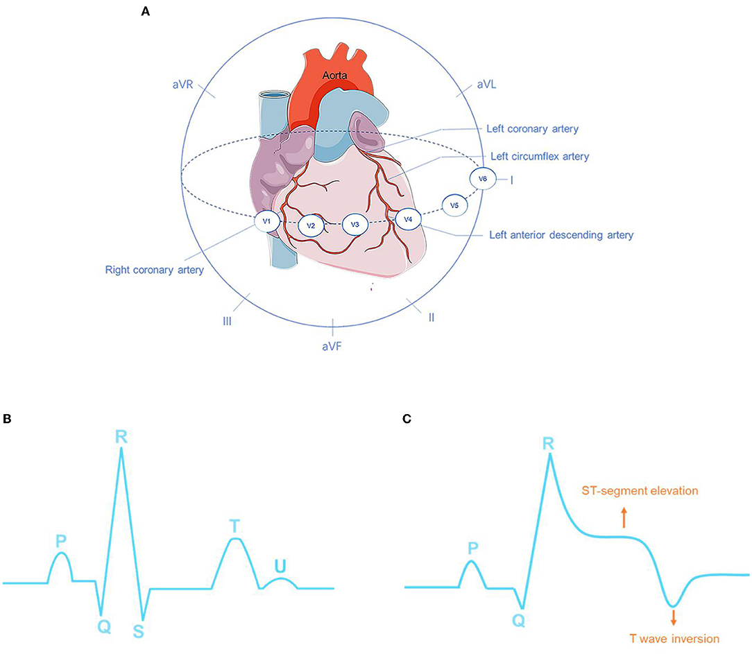
Frontiers Deep Learning For Detecting And Locating Myocardial Infarction By Electrocardiogram A Literature Review Cardiovascular Medicine
The Hidden Waves In The Ecg Uncovered Revealing A Sound Automated Interpretation Method Scientific Reports
Ecg Ekg Components

Electrocardiography Bioninja

A Typical Cycle Reflecting A Heart Beat In The Ecg Waveform Source Download Scientific Diagram

Collarbone Heartbeat Tattoo Do Want Latidos De Corazon Palabras Tatuajes Tatuajes De Linea
![]()
884 Beating Heart Stock Vector Illustration And Royalty Free Beating Heart Clipart
How Is The Heart Rate Determined On Electrocardiography Ecg

Ecg For One Normal Heartbeat Showing The Amplitudes And Time Durations Download Scientific Diagram

The Structure Of Normal Ecg Heartbeat Download Scientific Diagram
Automated Arrhythmia Detection From Electrocardiogram Signal Using Stacked Restricted Boltzmann Machine Model Springerlink
The Structure Of Normal Ecg Heartbeat Download Scientific Diagram

Ecg Rate Interpretation Litfl Medical Blog Ecg Library Basics
Comments
Post a Comment